Book of the Month: William Smellie's Sett of Anatomical Tables
The book I have chosen as this month’s book of the month is William Smellie’s A Sett of Anatomical Tables; a beautifully illustrated book of anatomical drawings, published in 1754 on the practice of midwifery. . It is an extremely rare text as only one hundred copies were produced at the time; the plates are especially fine and are far superior to any that had appeared before.[1]
 |
| Eight Table: The Uterus and the Internal Parts in the Sixth or Seventh Month of Pregnancy |
William Smellie (1741–44) was a Scottish obstetrician and one of the first notable man-midwives in Great Britain, regarded today as one of the greatest figures in obstetrical history. He was an exceptional teacher and the first to outline safe rules for the use of forceps. Smellie realised his contribution to the field of obstetrics and midwifery and states in the preface of his book:
I hope [...] that I have done something towards reducing that Art, into a more simple and mechanical method has hitherto been done. […], the following Tables [...] illustrate what I have taught and written on the subject.
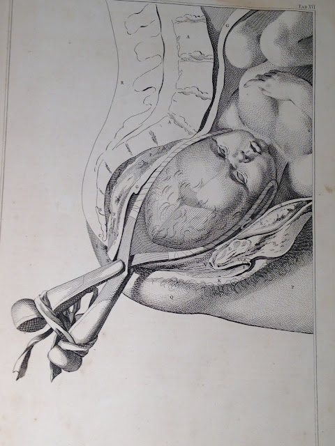
His most important contributions besides publishing his own teaching and describing the mechanism of labour, include the invention of ‘a machine’ (an obstetrical manikin for instructions) , and designing his own version of obstetrical forceps used to assist the delivery of a baby.
 |
| Sixteenth Table: The Head of the Baby is Helped Along with the Forceps |
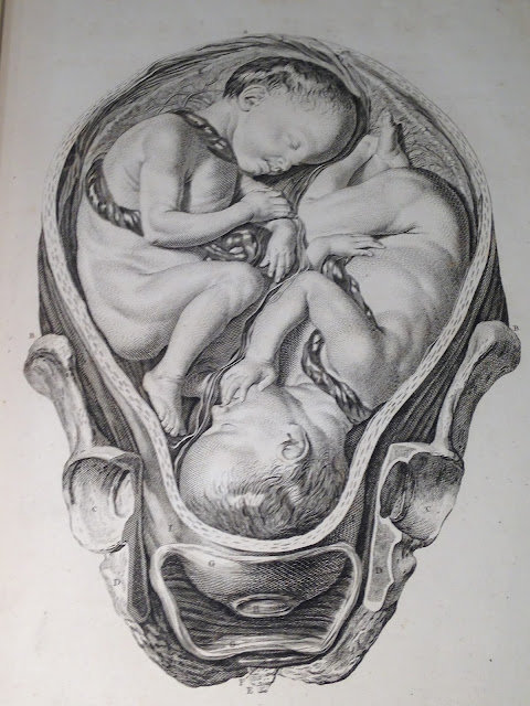
The plates were drawn mainly by Jan Van Rymsdyk with the addition of eleven by Petrus Camper (1722-1789), a Dutch physician, anatomist and midwife, and two by ‘another hand’. It is an extremely rare text as only one hundred copies were produced at the time; the plates are far superior to any that appeared before. The original sketches in mixed wet and dry red chalk, and later engraved by Charles Grignion, illustrate the most accurate depictions of childbirth at the time of publication. The illustrations are almost photo-realistic reproductions down to the tiniest wrinkle and hair. Not much is known about the Dutch artist, Jan van Rymsdyk, beyond that he lived and worked in England between 1745 and 1780. Jan van Rymsdyk’s first assignment as a medical illustrator seems to be for William Smellie’s Sett of Anatomical Tables, published in 1754. It was probably because of this assignment that William Hunter (1718-1783), the Scottish anatomist and physician (and a pupil of Smellie), commissioned Van Rymsdyk to illustrate his great book on obstetrics, The Anatomy of the Human Gravid Uterus, published in 1774.
 |
| Tenth Table: Uterus at the Beginning of Labour |
When researching into art and medicine for this blog post, I went as far back as the High Renaissance. The study of anatomy during that time was obligatory for many schools of art; Italian Renaissance artists became anatomists by necessity.[2] Michelangelo (1475-1564) and Leonardo da Vinci (1452-1519), both made important scientific discoveries and their anatomical sketches are important insights into the functioning of the human body. The study of anatomy and the dissection of human bodies were quite different in the eighteenth century to the sixteenth and seventeenth centuries; there was a shift from dissecting whole bodies to just parts of the human anatomy.
RCPI’s copy of Smellie’s ‘A Sett of Anatomical Tables’ forms part of Fleetwood Churchill collection, Churchill was a leading obstetrician in Dublin in the mid-nineteenth century and donated an important library of obstetrical books to RCPI. The volume was adopted by Dr Mary Holohan, FRCPI, in 2013 under our Adopt A Treasure programme, which allowed essential conservation work to be carried out on it.