Background
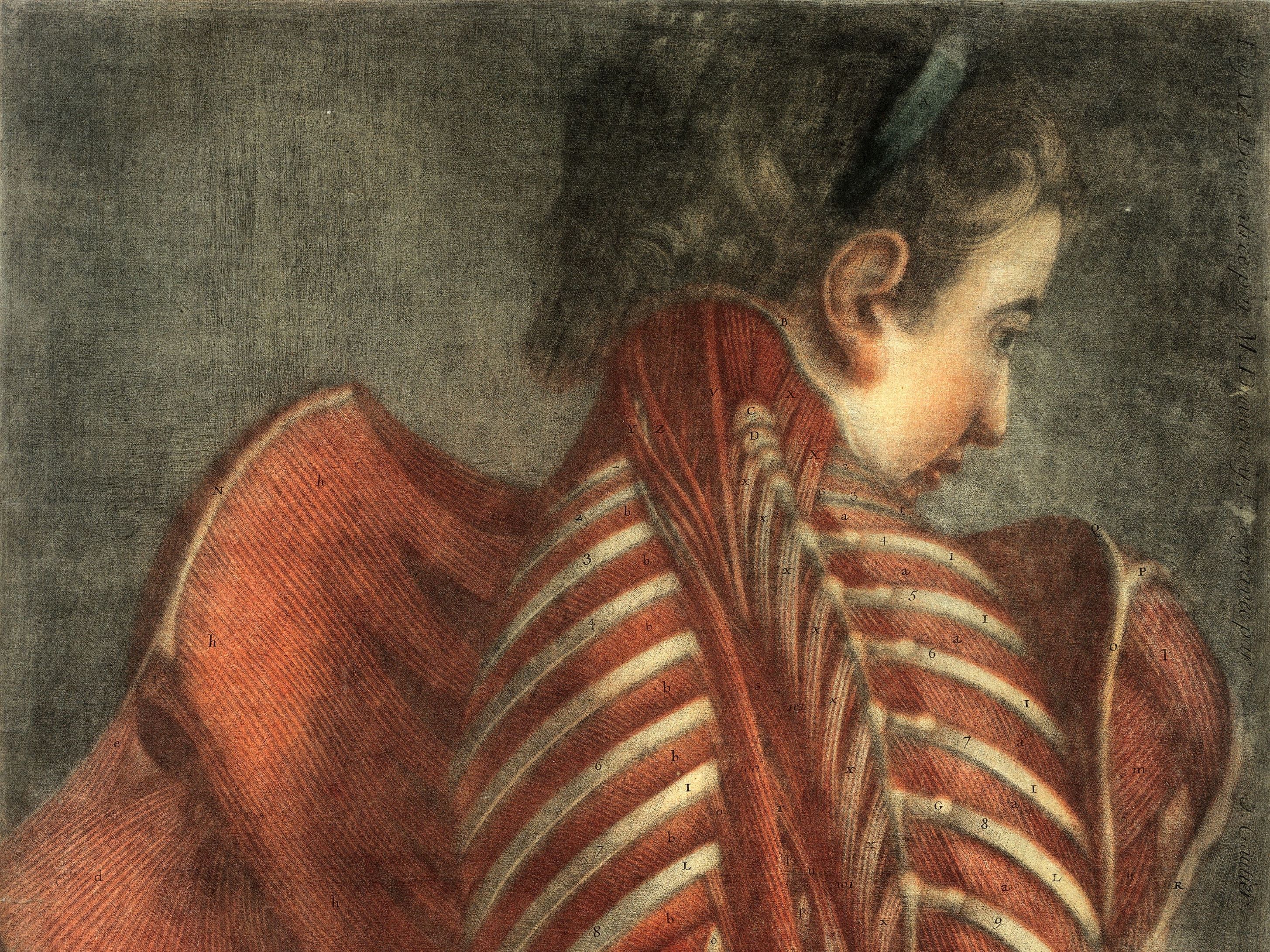
 From d'Agoty's Anatomie des parties de la generation de l'homme et de la femme (Paris, 1773)
Anatomical and descriptive illustration has played an important role in medical education and scholarship since antiquity.
From the skeleton, musculature, arteries, veins, nerves and viscera, the visual depiction of the human body traces the broad development of artistic practice and is found in finely illustrated manuscripts dating to the 4th and 5th centuries BCE.
This is mirrored in the development of printing from the sixteenth century. Colour was added to etched illustrations through polychrome woodcuts and hand tinted mezzotint, and from the nineteenth century though lithography and later, photography.
In contrast to this printing history, medical illustrations in the RCPI collection are single sheet watercolour illustrations and pencil sketches. Each illustration signed by the artist and inscribed with information on the patient depicted.
Likely requested by the attending physician, these were probably used in a teaching setting – introducing the medical student to the presentation of an ailment – and as illustrations to published articles.